Mitochondria Structure and Function With Diagrams

Mitochondria are one of the most important organelles of most eukaryotic cells. Within each mitochondrion, important processes are carried out in which different proteins, molecules, channels and membranes are involved. The functions of mitochondria are so advanced, it has been postulated that these organelles originated from a prokaryotic cell that engulfed an aerobic bacterium millions of years ago.
In this thedailyECO article, we the mitochondria structure and function. We also provide diagrams to give you a better idea of this structure.
What are mitochondria?
Mitochondria are highly specialized organelles found in the cytoplasm of most eukaryotic cells. These calls belong to animals, plants, fungi and protists. Although the vast majority of cells with a nucleus (eukaryotic cells) have mitochondria, there have been cells discovered which do not[1]. Although it is possible a eukaryotic cell can survive without mitochondria, it is believed even these cells had the organelle at one stage.
You can learn more about these different types of cells with our comparison article on the difference between eukaryotic and prokaryotic cells.
The mitochondria are small units within the cell that carry out specific functions for their development. They are associated with the cell membrane with the help of a double membrane, a feature represented in the diagram above. The shape of this organelle can be different depending on the type of cell in question. Some are in the shape of rods, long filaments or granules.
The number of mitochondria varies according to their cell type. For example, there are cells or tissues that have a much greater energy demand, such as muscle, brain or liver.
If you want to know more about cellular organelles, take a look at our related article on the function and structure of different cell organelles.
Mitochondria function
The function of the mitochondria is incredibly varied precisely because it carries out many vital functions within the cell. Of these various functions, there are two which are most closely associated with this type of cell organelle.
The main function of mitochondria as a cell organelle is cellular respiration through the use of oxygen. In addition, they are responsible for the production of chemical energy necessary for the cell to carry out its biochemical reactions. We can look at these two functions of mitochondria in more detail:
- Cellular respiration: there are different types of cellular respiration, all of which oxidize biological fuels to create energy. Cellular respiration which uses oxygen itself for this type of oxidization is known as aerobic respiration (anaerobic respiration uses electron acceptors that are not oxygen). Aerobic respiration almost always occurs in the mitochondria. Examples of these types of cell respiration include the Krebs cycle (a metabolic pathway in which energy is released by the oxidation of acetyl coenzyme A), oxidative phosphorylation and the electron transport chain (see diagram below).
- Chemical energy production: it is stored in the form of adenosine triphosphate (ATP), since this phosphate generates a high-energy bond. This ATP is catalyzed with the help of the transmembrane enzyme ATP synthase. This occurs thanks to the oxidation of amino acids, fatty acids and sugars, and this is what is known as the previously mentioned oxidative phosphorylation.
In the inner membrane of the mitochondria there are enzyme complexes composed of several proteins that have multiple activities:
- Use of molecular oxygen: often as an electron acceptor.
- Reduction and oxidation of different organic compounds: in this way a chain is formed through which electrons are transported.
- Proton pumping: towards the mitochondrial intermembrane space.
These respiratory chain complexes are divided into complex I (NADH dehydrogenase), complex II (succinate dehydrogenase), complex III (coenzyme Q-cytochrome c reductase) and complex IV (cytochrome oxidase). Learn more about how other organelles work with our article on the structure and function of chloroplasts.

The structure of mitochondria and their functions
Although the structure of the mitochondria can vary, there are some parts which are commonly used in their structure. The following are the main parts of mitochondria:
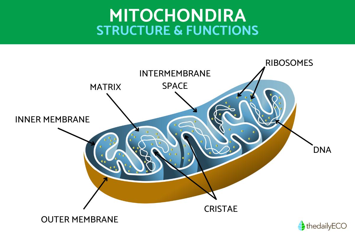
Outer membrane
The first part of the mitochondria that we see is the outer membrane. This both allows them to be delimited from other organelles, but its nature means they can communicate with these other parts. Mitochondria receive a lot of information from other parts of the cell. They do so using proteins known as porins. Porins are proteins with holes which allow the passage of ions and smaller proteins into the intermembrane space. All the proteins that enter must be unfolded, which happens thanks to the chaperone proteins. They also use protein complexes to introduce proteins.
Inner membrane
The next part of the mitochondria is the inner membrane that forms something known as the matrix (see diagram in main picture). It is analogous to the cytoplasm of the cell. Energy in the form of ATP comes from this region. Here metabolic certain processes occur, such as:
- Oxidative Phosphorylation
- ATP production
- Krebs cycle
- Pyruvate oxidization
- Amino acid oxidation
- Fatty acid oxidation
It also has a structural function because it is here where the other microorganelles of the mitochondria are located. These include ribosomes, DNA, ions and metabolites. The inner membrane is made up of a lipid bilayer where there are enzyme complexes made up of different proteins necessary for the electron transport chain.
Cristae
The outer and inner membranes are parts of the mitochondria that fold to form cristae. They are located mainly at the borders of the mitochondria, but delimited on the outside by the outer membrane. They fit perpendicular to the boundary of the mitochondria. It is in the membranes of these crista folds that the most important functions of mitochondria occur:
- Electron transport: with the help of enzyme complexes that transfer electrons from one place to another.
- Oxidative phosphorylation: as described above
- Stops electron blockages: by making electrons compact and maximizing their transfer.
Mitochondrial intermembrane space
Between the inner and outer membrane there is a space called the mitochondrial intermembrane space which is of vital importance for cell activity. It has a high content of enzymes necessary for respiration. Its main function is the reception of protons from the pumping of the enzymatic complexes.
The mitochondrial intermembrane space is of watery consistency. Enzymes and proteins that assist in the cellular process can be found here. Translocation also occurs. This is a process where mitochondrial matrix proteins are transported from outside the mitochondria. Lastly, they also transport fatty acids.
Mitoribosomes
Mitochondria also have ribosomes, called mitoribosomes or mitochondrial ribosomes. They have the same function as other ribosomes, i.e. to synthesize proteins through gene translation. They receive the information in the form of RNA to translate it into DNA. Learn more with our article on the difference between DNA and RNA.
Mitogenome (mtDNA)
Mitochondria have their own DNA. In fact, they are the only organelle with their own particular DNA. It always works in conjunction with the DNA of the nucleus to achieve coordinated activities. It has a small and circular shape. This mitochondrial DNA is only inherited from the mother, not from the father. It also cannot be passed on by genetic recombination.
In this mitochondrial DNA, genetic faults can occur with pathogenic results such as the development of Parkinson's disease. This is due to its proximity to oxidative metabolism. It also lacks protective histones, unlike the DNA of the cell nucleus, which does.
This DNA has been of great evolutionary interest. It has been explained by the principles of the endosymbiosis theory. This theory claims a prokaryotic cell engulfed an aerobically respiring bacterium to obtain its organelles, creating a eukaryotic cell and a symbiotic relationship. In this context, it is understandable the mitochondria is a special organelle with several micro organelles within it, acting as if it were a small cell by itself.
Learn more about how organisms can help each other with our article on the definition of symbiosis in biology with examples.
If you want to read similar articles to Mitochondria Structure and Function With Diagrams, we recommend you visit our Biology category.
1. Karnkowska, A., Vacek, V., Zubáčová, Z., Treitli, S. C., Petrželková, R., Eme, L., Novák, L., Žárský, V., Barlow, L. D., Herman, E. K., Soukal, P., Hroudová, M., Doležal, P., Stairs, C. W., Roger, A. J., Eliáš, M., Dacks, J. B., Vlček, Č., & Hampl, V. (2016). A Eukaryote without a Mitochondrial Organelle. Current biology: CB, 26(10), 1274–1284.
https://doi.org/10.1016/j.cub.2016.03.053
- Gómez-Pompa, A., Barrera, A., Gutiérrez-Vázquez, J., & Halffter, G. (1980). Biology: Unity, Diversity and Continuity of Living Beings. Mexico City: National Council for the Teaching of Biology.
- Medawar, P., & Medawar, J. (1988). From Aristotle to Zoos: A Philosophical Dictionary of Biology. Federal District: Economic Culture Fund.
- Gahl, W. (2018). Mitochondria. Available at: https://www.genome.gov/genetics-glossary
- Mendez, O. and Muhlia, A. (2018). Mitochondria, the ying-yang of life. Natural Resources and Society, 4(1), 12-21.


