Ribosomes Structure and Function in Biology

It can be helpful to look at cells as little factories. They have different departments and structures within them which carry out specific functions. Similar to a factory, these different structures within a cell work collectively to perform the overall function of the type of cell. Individually, they can only perform actions which their departments have the ability and resources to do. The individual structures within a cell are called organelles. Some organelles are common to all cells, but others are not. Ribosomes are a cell organelle which belong to the former category as they are found in all living cells.
Keeping with the factory analogy, ribosomes are considered macromolecular machines responsible for vital processes within living beings. thedailyECO discovers what these processes are by looking at the structure and function of ribosomes in biology and provides a summary and diagrams of them.
What are ribosomes in biology?
Ribosomes are cell organelles arranged in the cytoplasm within the cell. This is true of both eukaryotic and prokaryotic cells, meaning they are found in all types of cells. However, not all cells will have the same amount or types of ribosomes. For example, mammalian red blood cells do not have ribosomes in their mature form and the ribosomes in spermatozoa are very different from most other cells. This is because it was discovered mammalian ribosomes are found in the mitochondria of spermatozoa, instead of the cytoplasm[1].
Some of the common characteristics of ribosomes in biology are the following:
- They are globular in shape and do not have a membrane.
- They are part of the cellular machinery for making proteins, starting the process with one molecule of genetic information and translating it into another so that said information is materialized.
- Its composition is based on ribosomal RNA, hence its name. In addition, they are also composed of various ribosomal proteins. Learn more about this functionality of ribosomes by looking at our article on the difference between DNA and RNA.
With these characteristics in mind, we can say the following is a definition of ribosomes in biology:
Ribosomes are a type of organelle found in all cells that perform a biological synthesis of proteins from one type of molecule to another.
Ribosomes are essential for cellular function and survival. They ensure that the genetic code within DNA is accurately translated into proteins, which perform a multitude of roles within the cell. Without ribosomes, cells would be unable to produce the proteins necessary for life. Learn more about the structure of cells with our article on the definition and function of cell organelles.
Types of ribosomes
Ribosomes can be classified into several types. We can find different types of ribosomes depending on their arrangement within the cell, as well as the type of cell to which they belong. By looking at the following classification of ribosomes, we can provide a better summary of their function.
Types of ribosomes according to their location
The first classification responds to the arrangement of the ribosomes. Ribosome location will determine the location to which the processed proteins will be sent. For this reason, we can generally categorize ribosomes into the following:
- Attached ribosomes: these ribosomes are attached to the outside of the rough endoplasmic reticulum. They can also be found attached to the outer part of the nucellar membrane. Mitochondrial ribosomes are found attached to the mitochondrial membrane[2]. These produce proteins that will be inserted into membranes or which are used for secretion.
- Free ribosomes: they are free in the cytoplasm and can be individual or linked to RNA. When linked to RNA they will sandwich together to attract RNA molecules attached to amino acids, eventually forming a polypeptide chain after the amino acids are processed by the ribosomes.
Free ribosomes can be found in different areas. According to this characteristic, they can be classified as follows:
- Mitoribosomes: found free in the mitochondrial matrix, also known as mitochondrial ribosomes.
- Plastoribosomes: they are free in the stroma of the chloroplasts of prokaryotic cells, in plant cells or algae.
Types of ribosomes according to cell type
They can also be classified according to cell type in which they are located. In this case there are the following types of ribosomes:
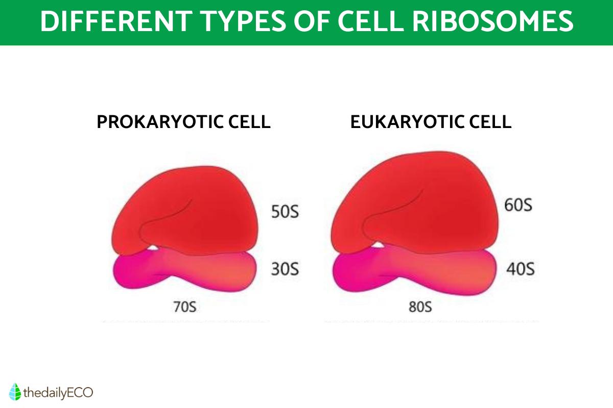
- Prokaryotic ribosomes: found in prokaryotic cells, i.e., cells without a true nucleus. They have a sedimentation level of 70S, smaller than eukaryotic ribosomes. We will go into more detail about this measure later. They are made up mostly of rRNA, and less than half is made up of protein.
- Eukaryotic ribosomes: the ribosomes that eukaryotic cells have, i.e., those with a nucleus. They have a higher sedimentation level than prokaryotic ribosomes, being 80S. More than half is made up of protein, and less than half is made up of rRNA. They are larger in size than prokaryotic ribosomes.
Understanding the differences between these types of ribosomes is crucial as it helps in the study of antibiotic functions and the development of treatments targeting bacterial infections. The unique characteristics of prokaryotic ribosomes, for instance, make them a target for antibiotics without affecting eukaryotic ribosomes. You can better visualize the difference between prokaryotic and eukaryotic ribosomes with the diagram below.
While the presence of a nucleus is one determining factor in the type of cell, you can learn more about the others with our article on the difference between eukaryotic and prokaryotic cells.

Function of ribosomes
Now we know about the different types of ribosomes, we can look at a better summary of their function and structure. Ribosomes are responsible for producing proteins and participate in the genetic control of the cell. For this, the ribosome used the following steps:
- Acquires and reads the messenger RNA information.
- Translates said information into a sequence of amino acids.
- The blocks that form the proteins are created which are not arranged randomly, but occur in a predetermined and ordered way.
There are organs that produce large amounts of protein, so the cells of their tissues need to have several ribosomes. An example of this is the pancreas, an organ that produces insulin.
Proper functionality of the ribosomes is essential for organisms to be able to fulfill protein processing. When there are ribosomal alterations, diseases such as Diamond-Blackfan anemia or dyskeratosis congenita can occur. These are congenital conditions which affect the integumentary and bone systems. Although they can occur in humans, they are considered rare.
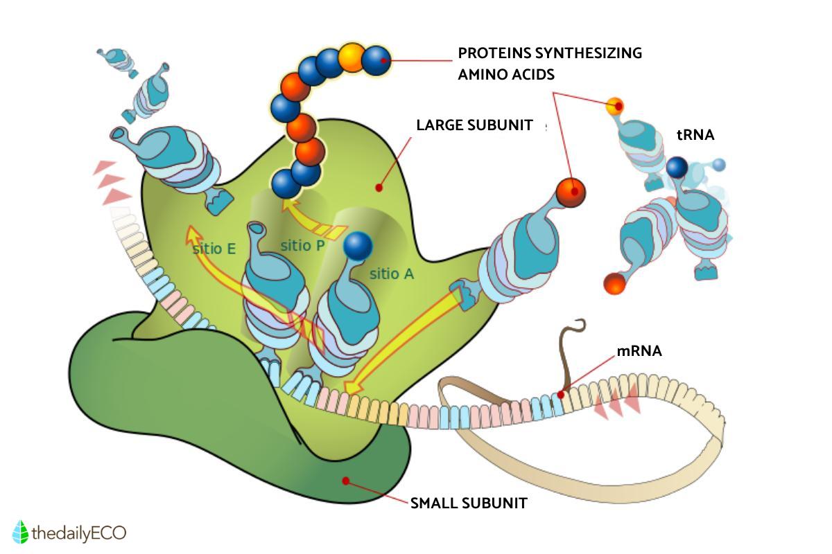
Ribosomes also play a critical role in cellular response to external stimuli. For instance, in response to stress or nutrient availability, ribosomes may adjust their protein synthesis rate, impacting the cell's adaptation to changing environments. This adaptability underscores the importance of ribosomes in maintaining cellular homeostasis. The diagram below presents the locations of these subunits during protein synthesis.

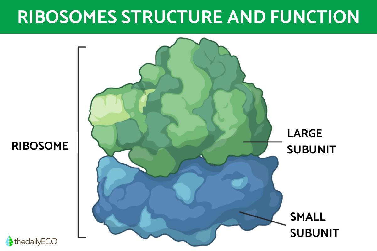
Ribosome structure
For the function of ribosomes to occur, they need to have a certain structure. Ribosomes are made up of two subunits that fit together, with each in turn made up of RNA and ribosomal proteins. The states of these two ribosomal subunits are the following:
- Large ribosomal subunit: has a central and two lateral bulges, with a front and a back.
- Small ribosomal subunit: the bulges on the large subunit help the small subunit to insert, the latter only having a head and a platform.
These subunits are measured with the sedimentation coefficient and are expressed in the Svedbergs unit, a non-SI metric unit which refers to the speed with which a particle settles during centrifugation. Depending on the type of cell in which the ribosome is found, this unit varies:
- Eukaryotic cells: the minor subunit is 40S and the major is 60S.
- Prokaryotic cells: the minor subunit is 30S and the major is 50S.
These subunits come together when they detect a messenger RNA (mRNA), positioning the small above the large one. The one which detects it is the small subunit, and then the large subunit mates.
With the help of transfer RNA (tRNA), it translates messenger RNA into a chain of amino acids that will form proteins. The small subunit is the one that links the tRNA to the mRNA. The large subunit allows the bonds that join the amino acids to form. The subunits separate when they detect a termination sequence, indicating that the protein is complete.
In the larger subunit there are three sites or clefts necessary for the transfer RNAs to insert and complete their translation process into proteins. Such sites are:
- Site A or amino acid: where the tRNA aminoacyl is inserted, which carries the first amino acid.
- P or peptide site: where the peptidyl tRNA is inserted, which leads to the chain of peptides and to the proteins as a result. You can see this process in the diagram below.
- Site E: is the final site, where the tRNA from the P site moves and replaces the tRNA that was at this site.
These newly created proteins are responsible for various functions within the organism, including enzymatic activity, structural support, and regulation of cellular processes. Understanding ribosome structure is essential for comprehending how genetic information is translated into functional proteins, a process fundamental to life. Discover one of their most vital uses by looking at the phases of the cell cycle.

If you want to read similar articles to Ribosomes Structure and Function in Biology, we recommend you visit our Biology category.
1. Gur, Y., & Breitbart, H. (2006). Mammalian sperm translate nuclear-encoded proteins by mitochondrial-type ribosomes. Genes & development, 20(4), 411–416.
https://doi.org/10.1101/gad.367606
Greber, B. J., & Ban, N. (2016). Structure and Function of the Mitochondrial Ribosome. Annual review of biochemistry, 85, 103–132.
https://doi.org/10.1146/annurev-biochem-060815-014343
- Campbell, N., & Reece, J. (2006). Biology. Madrid: Panamerican Medical Editorial.
- Marcey, D. (2014). Introduction to the structure of the ribosome. Retrieved from: http://biomodel.uah.es/model1j/rna-prot/ribosoma.htm
- Miguel Hernandez University. (2006). General organization of cells: Cytosol and endomembrane system. Retrieved from: http://retina.umh.es/docencia/confsvivos/temas/Tema_11_Ribosomas.pdf


