Cilia vs. Flagella Differences in Function and Structure

Cilia and flagella are specialized structures located on the surface of single-celled eukaryotic cells, as well as organisms such as bacteria and archaea. While they have differences in terms of structure and function, they are often confused with each other due to their similarities. In terms of structure, they both emerge from a basal body and are made from microtubules which are elongated and facilitate movement. In terms of function, they share similarities in terms of cellular movement, sensory detection and signal transduction. Although they share various traits, it is important to know the cilia vs. flagella differences in function and structure. thedailyECO explains these differences and provides diagrams to help visualize them.
What are cilia?
Cilia are specialized cellular structures found on the surface of many eukaryotic cells. They are considered types of cell organelle and are linked to the cell membrane. They are small, mobile projections similar to hairs or eyelashes that protrude from the plasma membrane itself. In this way, cilia are similar to trichomes which are hair-like appendages on plants, algae and other organisms.
Function of cilia
Cilia perform several important functions in certain cells. Below are some of the main functions of cilia along with examples:
- Fluid movement and transport: cilia present on respiratory epithelial cells coordinate to move mucus toward the throat, helping to remove foreign particles and protect the lungs.
- Sensory detection: in the sensory cells of the inner ear, cilia capture sound vibrations and convert them into electrical signals that the brain can interpret as sound.
- Signal transduction: cilia can transduce signals from outside the cell into intracellular responses. For example, cilia in kidney cells are sensitive to fluid flow and can detect changes in pressure. This triggers a cellular response that regulates the reabsorption of water and salts in the kidney.
Structure of cilia
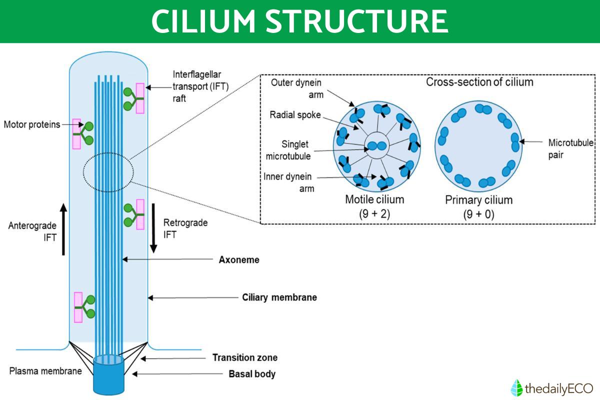
Cilia are primarily composed of microtubules. These are tubular protein filaments that form an internal skeleton called the axoneme. The axoneme of the cilia consists of a microtubule arrangement called 9+2 which consists of nine pairs of peripheral microtubules surrounding two central microtubules.
The 9+2 structure is characteristic of motile cilia which are found in cells such as respiratory tract epithelial cells and renal tubular cells. These mobile cilia have a coordinated and rhythmic movement that helps the movement of fluids or particles along the cell surface. For example, cilia on cells in the respiratory tract help move mucus and trap foreign particles for later elimination.
There are also primary cilia which have a 9+0 structure, meaning they have nine pairs of peripheral microtubules, but lack the central microtubules. These primary cilia are less motile and perform various other functions, such as sensing environmental signals and transducing chemical and mechanical signals.

What are flagella?
Flagella are another type of specialized cellular structure found in some plant and animal cells, as well organisms, such as bacteria, archaea and unicellular eukaryotes. They are elongated, mobile appendages that protrude from the cell surface and are involved in cellular locomotion. They are considered motile organelles because they are capable of locomotion within the cell or its surrounding environment.
Unlike cilia, which are usually numerous and short, a flagellum consists of a single, long and whip-shaped structure. However, there can be multiple flagella on the same cell, but it will depend on the type of cell and its function. They are composed mainly of proteins, such as flagellin in bacteria and tubulin in unicellular eukaryotes.
Function of flagella
The main function of flagella is to allow cell movement, especially locomotion in liquid media. These specific functions include:
- Motile bacteria: many bacteria have flagella that allow them to move toward or away from certain chemical or physical stimuli. For example, flagellated bacteria such as Escherichia coli can use their flagella to swim in search of nutrients or to move away from antibacterial substances.
- Flagellated protists: unicellular protists, such as protozoa, often have one or more flagella that allow them to move in their aqueous environment. For example, paramecium is a ciliate protozoan that has a flagellum to help it move quickly and efficiently in water.
- Flagellated algae: the green algae Chlamydomonas reinhardtii has two flagella that allow it to swim and orient itself towards light to carry out photosynthesis.
- Flagellated sperm cells: certain multicellular organisms have sperm cells with a single flagellum which is used to swim towards the
Structure of the flagella
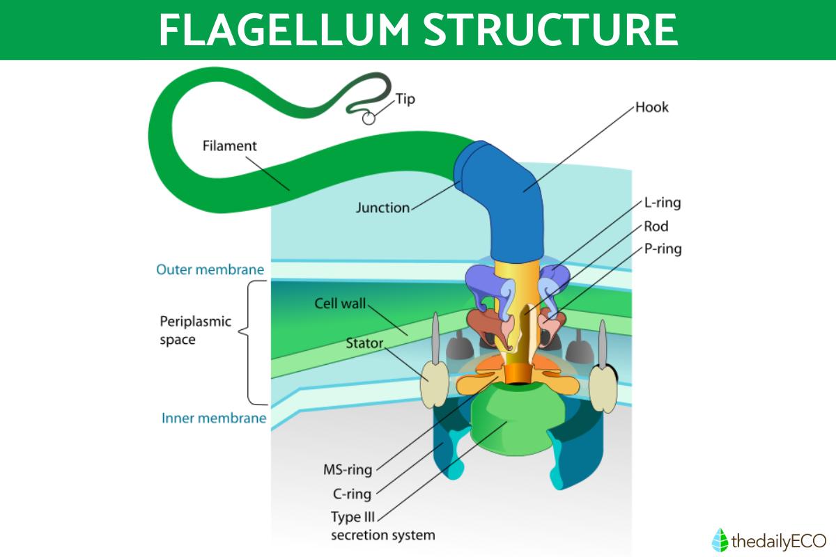
Flagella have a basic structure consisting of three main components:
- Filament: it is the external part of the flagellum and consists of a helical chain of flagellin subunits, which provides the structure and the ability to move.
- Hook: it is a flexible joint that connects the filament with the basal body. It allows the flagellum to move in different directions.
- Basal body: it is the base of the flagellum found inside the cell. It contains the rings and the flagellar motor, which are responsible for generating the energy necessary for the movement of the flagellum.
In unicellular eukaryotic organisms, such as protozoa and some algae, flagella have a structure similar to the 9+2 axoneme of cilia. They consist of nine pairs of peripheral microtubules and two central microtubules. These flagella are capable of moving in an undulating or flapping manner, propelling the organism through an aqueous medium.
Learn about the differences between eukaryotes and prokaryotes in our related guide.

What are the dDifferences between cilia vs. flagella
There are several differences between cilia and flagella. Some of the main differences between these two cellular structures are the following:
- Structure and length: cilia are shorter their structures are more numerous, while flagella are longer and generally present in smaller quantities. Cilia are usually between 2 and 10 micrometers in length, while flagella can reach several micrometers or even millimeters in length.
- Distribution pattern: cilia are usually arranged in rows along the cell surface, while flagella are usually single structures that protrude from the cell.
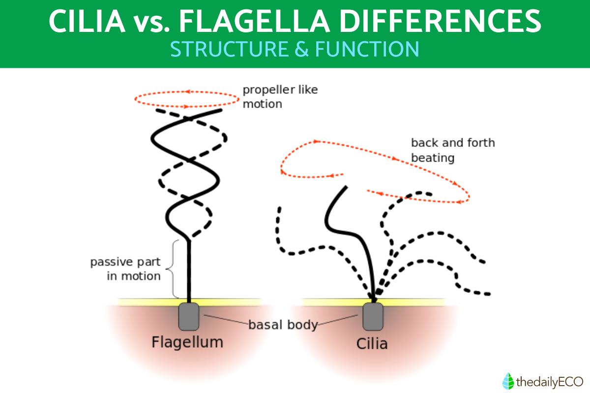
- Movement: cilia generally make coordinated, rhythmic, beating movements that allow the generation of directional flow. Instead, flagella can move in an undulating or whip-like manner to generate movement and propulsion.
Now you know about what cilia vs. flagella are, you may want to learn more about other types of cellular structures. You can do so with our articles explaining what is cytoplasm and what are ribosomes?
If you want to read similar articles to Cilia vs. Flagella Differences in Function and Structure, we recommend you visit our Biology category.
- Campbell, N. A., & Reece, J. B. (2007). Biology. Argentina: Editorial Médica Panamericana S. A.


